
See more, treat precisely
The magnified view shows canal openings and internal conditions during treatment, helping find hidden canals and remaining inflammation.
Root Canal
Tooth nerves run thin and deep where the naked eye cannot follow. We work under a dental operating microscope, examining the canals under magnification — and before any tooth is extracted, we first ask whether it can be saved.
 GENERAL CARE
 Root Canal
When decay or trauma inflames the nerve inside a tooth, the inflamed tissue is removed and the empty canals are disinfected and sealed so the tooth itself can be kept. Root canal treatment is the last step that preserves a natural tooth instead of extracting it; afterward, a crown or other final restoration usually protects the tooth from chewing forces.
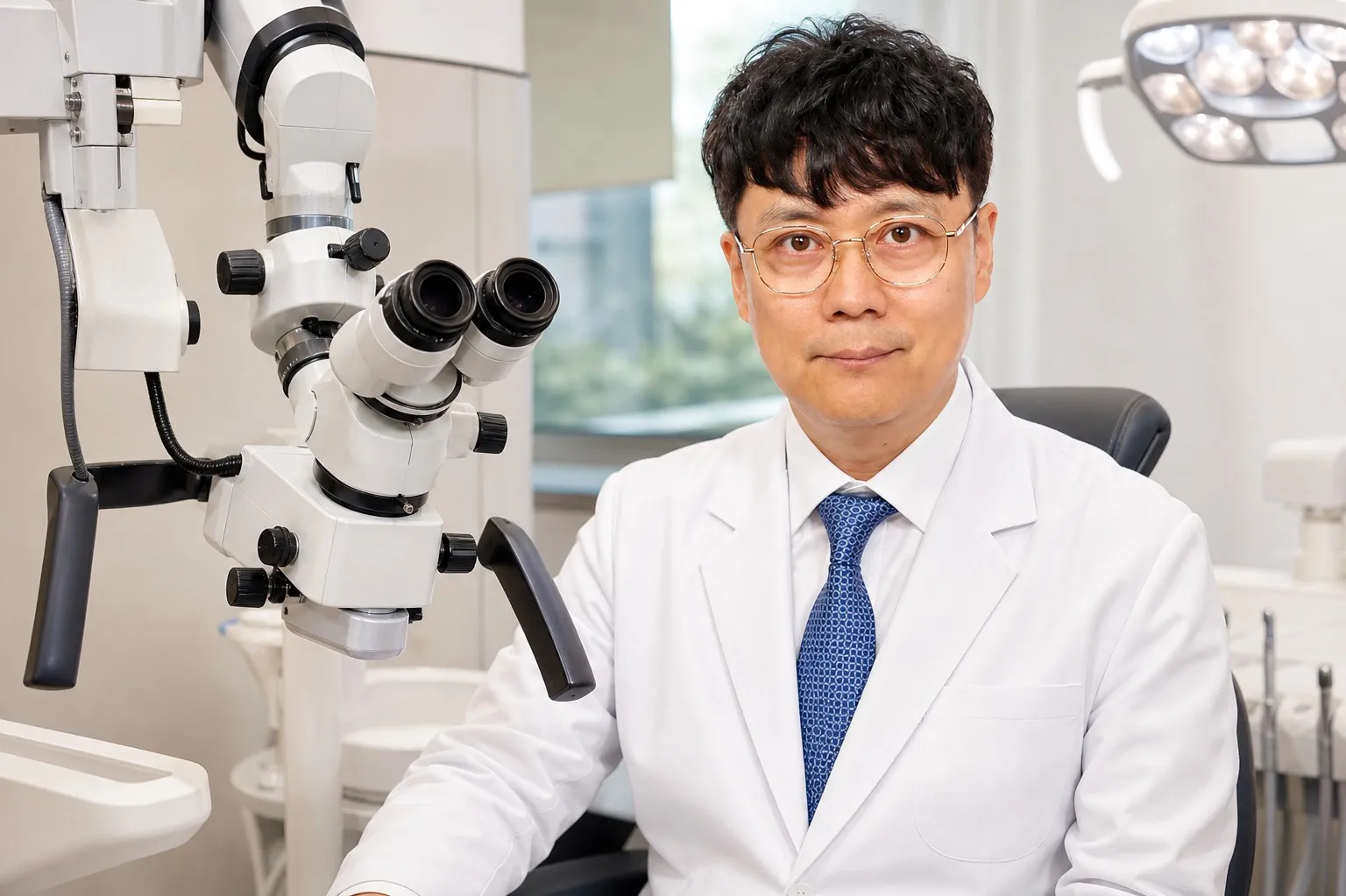
Microscope
A dental operating microscope magnifies the treatment field up to 25×, helping locate fine canals and hairline cracks the naked eye can miss.
The magnified view shows canal openings and internal conditions during treatment, helping find hidden canals and remaining inflammation.
When X-rays alone cannot explain pain, magnification can reveal cracks or subtle lesions and help reach the cause.
Blocked canals, curved canals, and retreatment after a previous failure can be approached with a precise plan.
Process
The number of visits varies with the inflammation and canal anatomy.
Access opening
A small opening is made in the tooth to reach the inflamed nerve tissue.
Cleaning and shaping
Inflamed tissue is removed and the canals are cleaned and disinfected.
Canal filling
The emptied canals are densely filled and sealed to lower the risk of reinfection.
Final restoration
The weakened tooth is protected from chewing forces with a crown or other final restoration, chosen by its position and remaining structure.
Indications
The situations below call for an evaluation of the nerve. Diagnosis is confirmed by examination and radiographs.

Whether a tooth needs extraction is judged from its remaining structure, cracks, periodontal support, and the extent of infection. When preservation is possible, root canal treatment or retreatment comes first — keeping a natural tooth comes before any replacement. For protecting a treated tooth, see our guide to all-ceramic crowns.
Quick Consult
Leave your number and we will call you back during clinic hours.
Request Received
We will contact you during clinic hours. For urgent matters, please call +82-2-3448-0028.
The available range depends on your condition. Feel free to ask us.
FAQ
Treatment is done under local anesthesia, so discomfort during the procedure is limited for most patients. Mild pressure or soreness between and after visits usually settles with time; increasing pain or swelling should be checked promptly.
Usually two to four visits depending on the inflammation and the number and shape of the canals. Stopping mid-treatment raises the risk of reinfection, so completing the crown restoration matters.
A tooth without its nerve can darken over time. Molars are covered by crowns, so this rarely shows; for visible front teeth, we discuss crowns or veneers to improve shade and shape together.
An implant cannot fully replace the ligament and sensation of a natural tooth. When a tooth can be preserved, saving it comes first; extraction and replacement are discussed only when preservation is judged unlikely.
This page is general information about the treatment. Diagnosis, treatment options, and duration vary by individual condition — an in-person examination is how we plan your actual care.
We find the cause of the pain, evaluate the nerve, and consider preservation first. Reception until 5:30 pm weekdays, 12:30 pm Saturdays.